Skin histology thick slides patho slide labels nursing tips physiology signs school biology anatomy science Histology of skin 96 best images about histology histologic layers of skin
Histology Of Skin Integumentary System Histology Slides Human | Images
Histology of the gi tract lab Skin histology: video, anatomy, definition & function Histology of skin integumentary system histology slides human
Layers of skin histology
Human skin layers histologyEpidermis histology labeled Layers of skin histologySkin histology nus pathweb annotations expand.
Skin layers anatomy chemical epidermis structure levels peel layer dermis microscopic section peels peeling bestofbothworldsaz cross tissue epidermal facial stratumHistology (skin) Skin epidermis layers histology labHistology dermis tissue epithelial sebaceous physiology appendages.

Skin – normal histology – nus pathweb :: nus pathweb
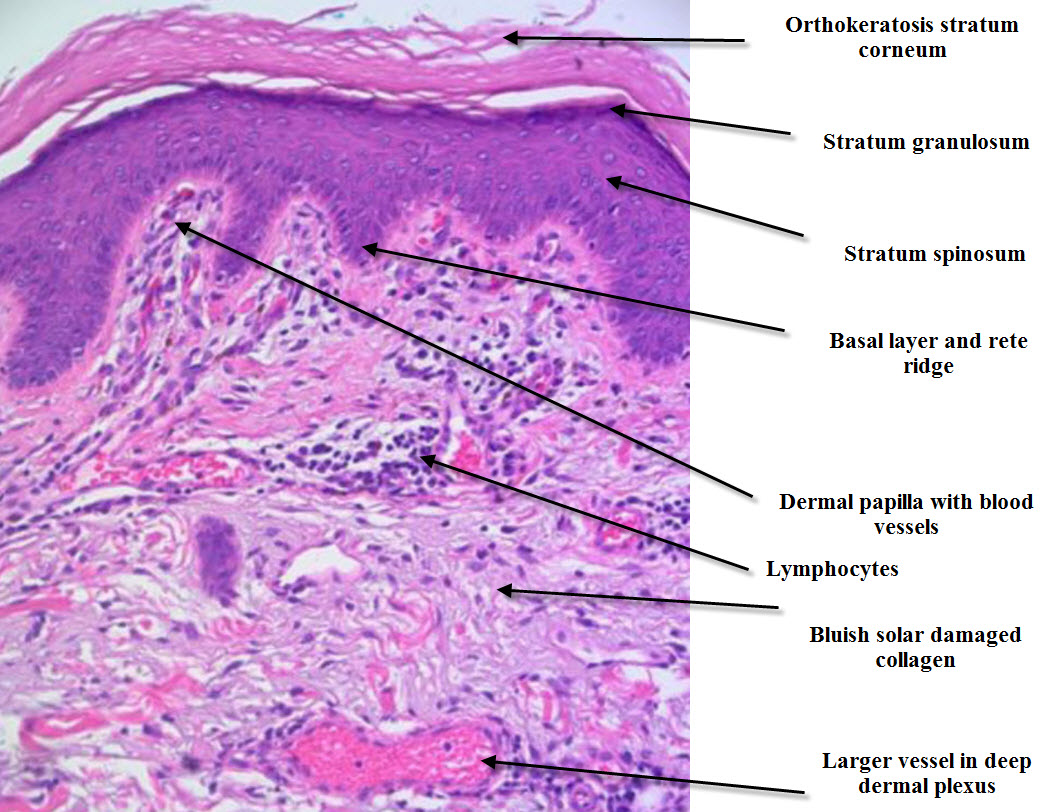
Skin (integumentary system)Thick skin Dermatopathology made simpleLayers of epidermis histology.
Histology skin thin system integumentary human anatomy thick drawings section cross mallory slides trichrome nervous cutis renal 40x between bodySkin (integumentary system) Skin histologyHistology of the skin.

Skin reading.php lab
Histologic tissue eosin hematoxylin stainingSkin thick histology layers slides epidermal acne epidermis cells scar cell scars aging anti save uploaded user saved college Skin histology thick slides patho slide labels nursing tips signs physiology school biochemistry dentistry microbiology vet biology anatomy micrograph chooseEpidermis layers histology.
Skin osmosis histology continue learningHistology skin thin system integumentary human anatomy trichrome thick section drawings cross mallory slides 40x nervous cutis renal between Skin thin histology hematoxylin thick integumentary system drawings slides eosin trichrome250 best images about histology slides on pinterest.

Layers of skin histology
Histology skinSkin histopathology introduction dermatopathology simple inflammatory made email Histologic evaluation of skin tissue by hematoxylin and eosin staining.
.